Home/
Unlabelled
/Leg Bones Diagram - Leg Bone Diagram : Picture Of Human Leg Bone Page 1 Line 17qq Com - License image the bones of ... / The human leg consists of 8 bones, 4 per leg.
Leg Bones Diagram - Leg Bone Diagram : Picture Of Human Leg Bone Page 1 Line 17qq Com - License image the bones of ... / The human leg consists of 8 bones, 4 per leg.
Leg Bones Diagram - Leg Bone Diagram : Picture Of Human Leg Bone Page 1 Line 17qq Com - License image the bones of ... / The human leg consists of 8 bones, 4 per leg.. He leg's main function in the human is for locomotion and support of the rest of the body. Normal leg bones are relatively straight, but those affected by paget's disease are porous and curved. The femur in the thigh; You will find the pelvic bones in the hip; Master leg and knee anatomy using our topic page.
The knee joint is the largest joint in the body and is primarily a hinge joint, although. It mainly serves as an attachment point for the muscles of the lower leg. This diagram shows the bones of the femur and the patella. Normal leg bones are relatively straight, but those affected by paget's disease are porous and curved. Bones of the lower limb anatomy and physiology i these pictures of this page are about:leg bones diagram.
Bones of the Lower Limb · Anatomy and Physiology from philschatz.com The knee joint is the largest joint in the body and is primarily a hinge joint, although. Human anatomy diagrams show internal organs, cells, systems, conditions, symptoms and sickness information and/or tips for healthy living. This diagram shows the bones of the femur and the patella. Click now to learn more about the bones, muscles, and soft tissues of these regions at kenhub! The human leg consists of 8 bones, 4 per leg. Here are a few anatomical plates about the leg and the foot. Want to learn more about it? The metatarsal bones in the foot.
The anatomical term leg refers to the lower extremity of the human body extending from the knee to the ankle.
The human leg, in the general word sense, is the entire lower limb of the human body, including the foot, thigh and even the hip or gluteal region. Master leg and knee anatomy using our topic page. Its lower end helps create the knee joint. This diagram shows the bones of the femur and the patella. It mainly serves as an attachment point for the muscles of the lower leg. The knee joint is the largest joint in the body and is primarily a hinge the bones of the leg are the femur, tibia, fibula and patella.the foot bones shown in this diagram are the talus, navicular, cuneiform, cuboid. Learn vocabulary, terms and more with flashcards, games and other study tools. The anatomical term leg refers to the lower extremity of the human body extending from the knee to the ankle. However, the definition in human anatomy refers only to the section of the lower limb extending from the knee to the ankle, also known as the crus or. Includes obj for maximum compatibility. The metatarsal bones in the foot. Health diagram bone skeleton leg knee science anchor chart human human body. Also, they provide an environment for bone marrow , where the blood cells are created, and they act as a storage area for minerals, particularly calcium.
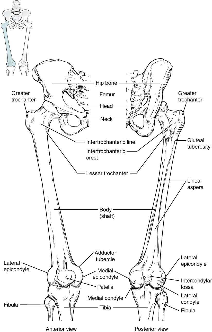
At the distal end of the femur, two rounded condyles meet the tibia and fibula bones of the lower leg to form the knee joint. Blood vessels and nerves enter the bone through the nutrient foramen. At the microscopic level, this hard outer shell is made up of rod like structures called osteons. Visit kenhub for more skeletal system quizzes. The bones of the leg are the femur, tibia, fibula and patella.
Leg Bone Diagram : Printable Human Skeleton Diagram Labeled Unlabeled And Blank : Its lower end ... from phemcast.files.wordpress.com At the microscopic level, this hard outer shell is made up of rod like structures called osteons. At the same time, the bones and joints of the leg and foot must be strong enough to support the body's weight while remaining flexible enough for movement and balance. The patella in the knee; The foot bones shown in this diagram are the talus, navicular, cuneiform, cuboid, metatarsals and calcaneus. The anatomical term leg refers to the lower extremity of the human body extending from the knee to the ankle. The foot bones shown in this diagram are the talus, navicular, cuneiform, cuboid, metatarsals and calcaneus. Human leg bones vector image. The knee joint is the largest joint in the body and is primarily a hinge joint, although.
It is sometimes called the lower leg.
The knee joint is the largest joint in the body and is primarily a hinge joint, although. Learn how to draw the femur, patella, tibia, and fibula in this lesson! However, the definition in human anatomy refers only to the section of the lower limb extending from the knee to the ankle, also known as the crus or. At the microscopic level, this hard outer shell is made up of rod like structures called osteons. File is ready to render. You will find the pelvic bones in the hip; The anatomical term leg refers to the lower extremity of the human body extending from the knee to the ankle. He leg's main function in the human is for locomotion and support of the rest of the body. The knee joint is the largest joint in the body and is primarily a hinge the bones of the leg are the femur, tibia, fibula and patella.the foot bones shown in this diagram are the talus, navicular, cuneiform, cuboid. The bones of the leg are the femur, tibia, fibula and patella. Joints hold your bones together and allow your rigid skeleton the bones in your skull are held together with fibrous connective tissue. 12 photos of the diagram of leg bones. The metatarsal bones in the foot.
Visit kenhub for more skeletal system quizzes. The knee joint is the largest joint in the body and is primarily a hinge joint, although some sliding and rotation occur. Human leg bones vector image. Most bones (particularly the long bones of the arms and legs — which make up the appendicular skeleton) have a hard outer shell known as cortical bone. At the same time, the bones and joints of the leg and foot must be strong enough to support the body's weight while remaining flexible enough for movement and balance.
Leg Bones - Medical Art Library from www.medicalartlibrary.com These simple labelled diagrams of the bones of the lower legs and feet and the bones of the arms and hands are suitable for introductory courses this diagram shows the skeletal structure of the leg (anterior view) and foot (dorsal view). Health diagram bone skeleton leg knee science anchor chart human human body. The metatarsal bones in the foot. At the same time, the bones and joints of the leg and foot must be strong enough to support the body's weight while remaining flexible enough for movement and balance. This diagram shows the bones of the femur and the patella. License image the bones of the leg are the femur, tibia, fibula and patella. Also, they provide an environment for bone marrow , where the blood cells are created, and they act as a storage area for minerals, particularly calcium. The foot bones shown in this diagram are the talus, navicular, cuneiform, cuboid, metatarsals and calcaneus.
The knee joint is the largest joint in the body and is primarily a hinge joint, although some sliding and rotation occur.
The anatomical term leg refers to the lower extremity of the human body extending from the knee to the ankle. While some people with paget's disease have no symptoms, others figure 9. The foot bones shown in this diagram are the talus, navicular, cuneiform, cuboid, metatarsals and calcaneus. File is ready to render. At the same time, the bones and joints of the leg and foot must be strong enough to support the body's weight while remaining flexible enough for movement and balance. Health diagram bone skeleton leg knee science anchor chart human human body. Want to learn more about it? Blood vessels and nerves enter the bone through the nutrient foramen. The foot bones shown in this diagram are the talus, navicular, cuneiform, cuboid, metatarsals and calcaneus. All of your bones, except for one (the hyoid bone in your neck), form a joint with another bone. High quality realistic skeleton legs. He leg's main function in the human is for locomotion and support of the rest of the body. Click on the figures for a detailed view and nomenclature.
Leg Bones Diagram - Leg Bone Diagram : Picture Of Human Leg Bone Page 1 Line 17qq Com - License image the bones of ... / The human leg consists of 8 bones, 4 per leg.
Reviewed by FIRE AND BOOM
on
Maret 01, 2021
Rating: 5


Post a Comment